Guide to photographing Procambarus simulans and P. curdi for identification
The two members of the Procambarus simulans complex, Procambarus simulans and
P. curdi , are readily identified as such by photos that are typically provided in iNaturalist
observations. Very few observations, however, provide the necessary photos needed to distinguish
them from each other. In certain geographic regions, the ranges are distinct and they can simply be
distinguished by range. These areas include the western halves of Texas and Oklahoma and parts of
SE Texas. The two occur together in NE Texas, SE Oklahoma, and parts of SE Texas. Very notably
they both occur in the Dallas/Fort Worth area. For these regions, specific photos must be provided
to allow identification to species. Otherwise the identification will have the be left at "Procambarus
simulans complex." The following guide describes the anatomical features and photo angles needed.
The two species, insofar as is currently known, are only identifiable by
features of the gonopod (male) or the annulus ventralis (female).
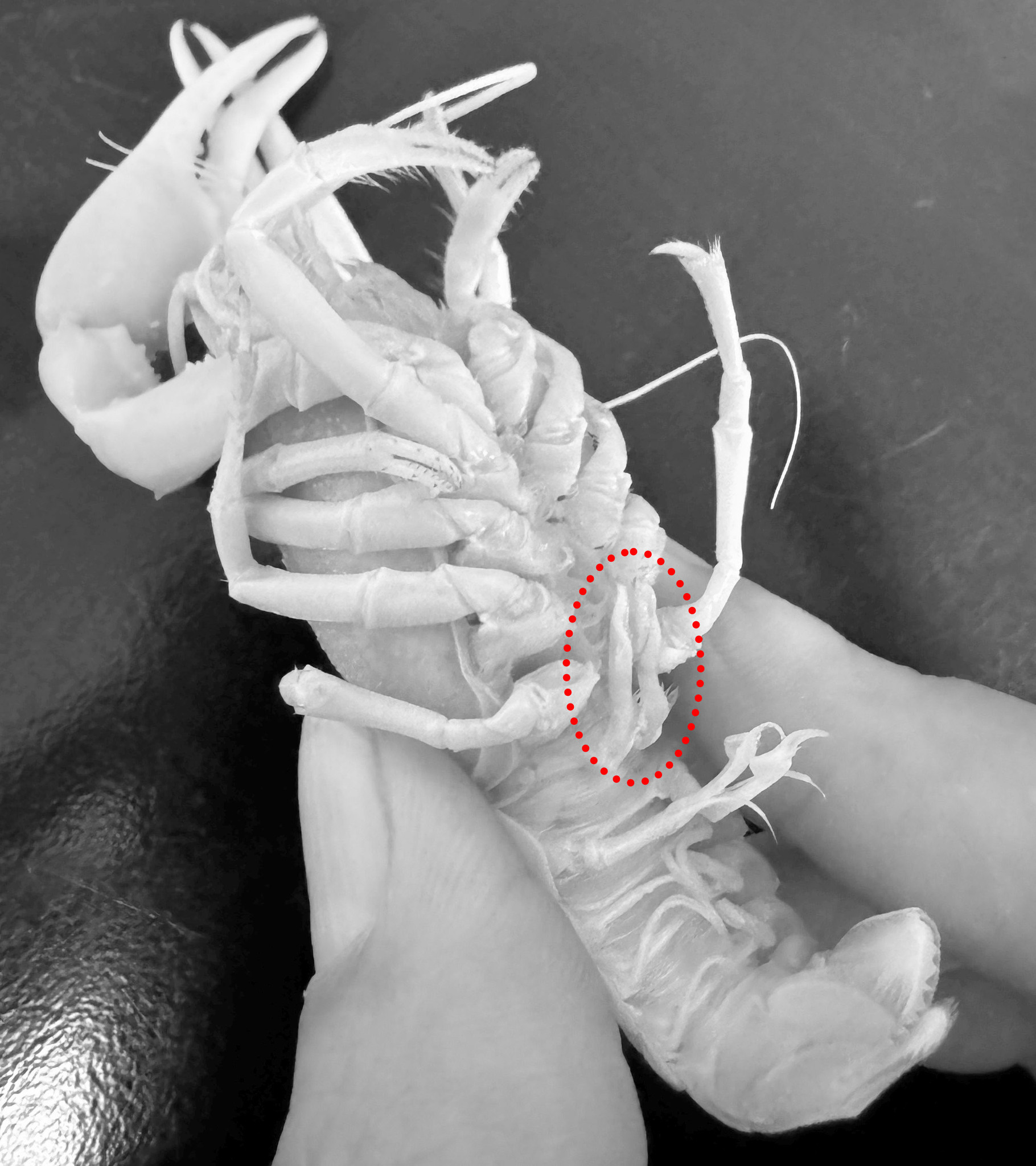
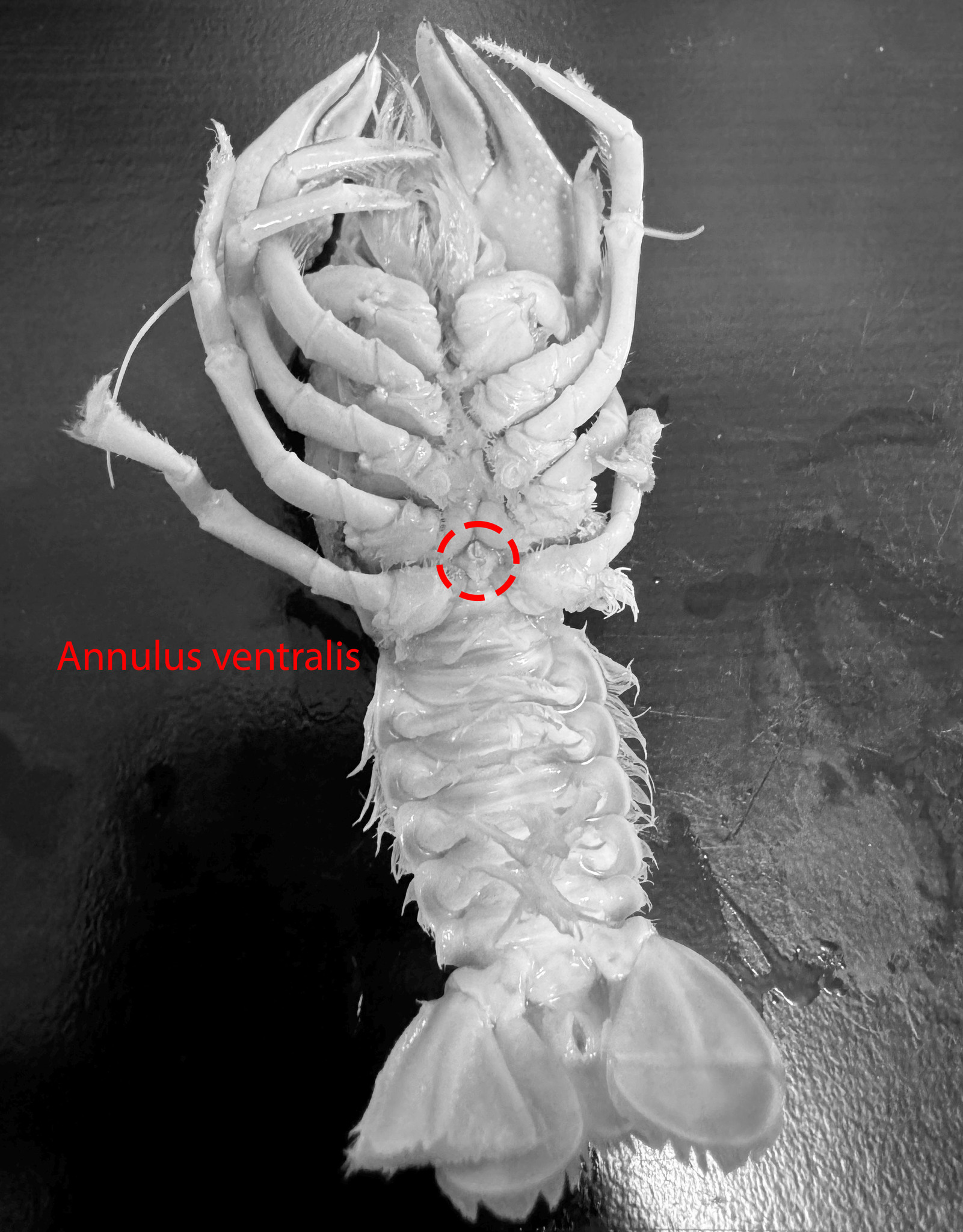
Below is a picture of the ventral surface of a Procambarus simulans male
with the pair of gonopods indicated by the dashed red circle. The photographs should
be taken at this angle with focus on the apex of it's left gonopod
(one on right in photo).
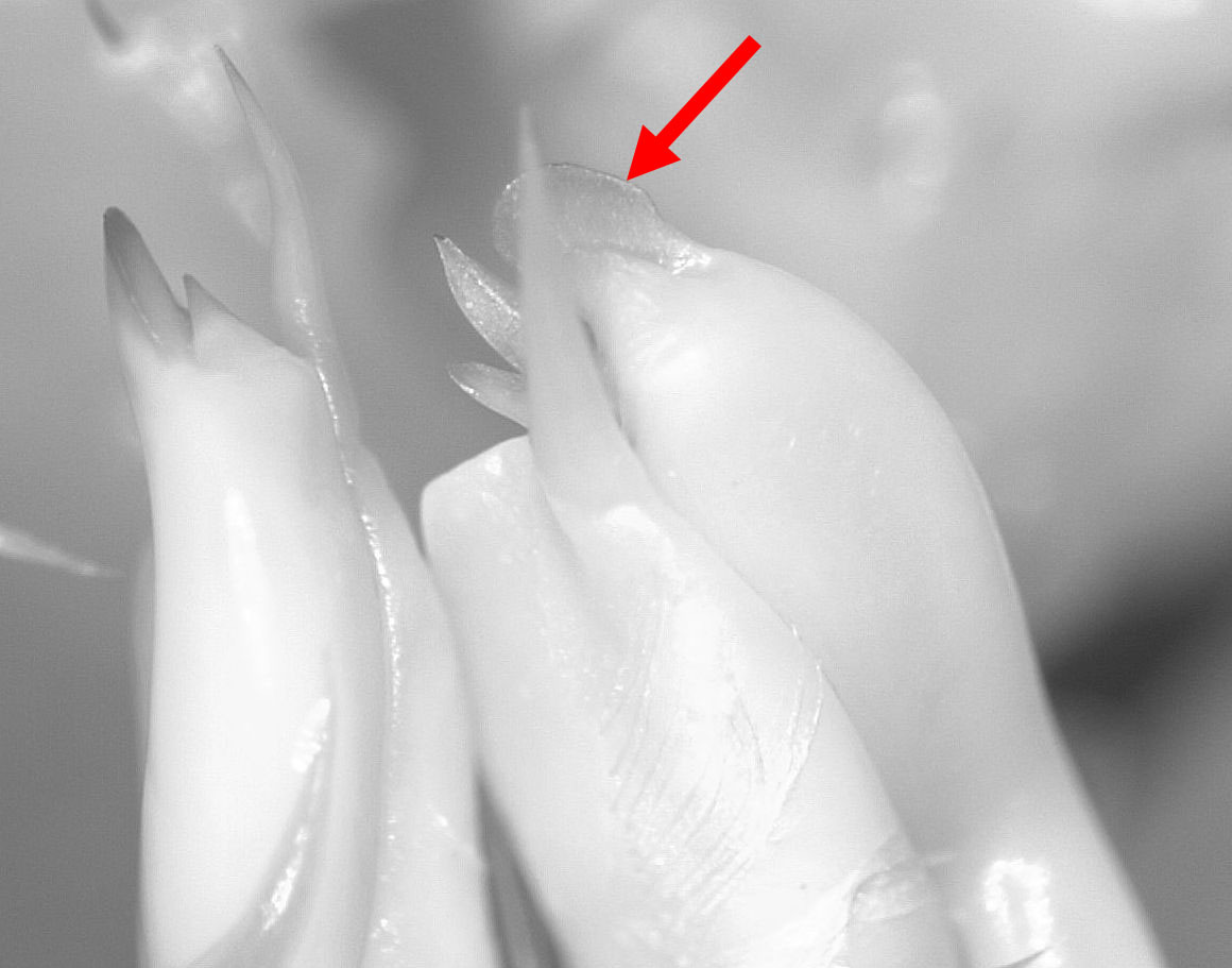
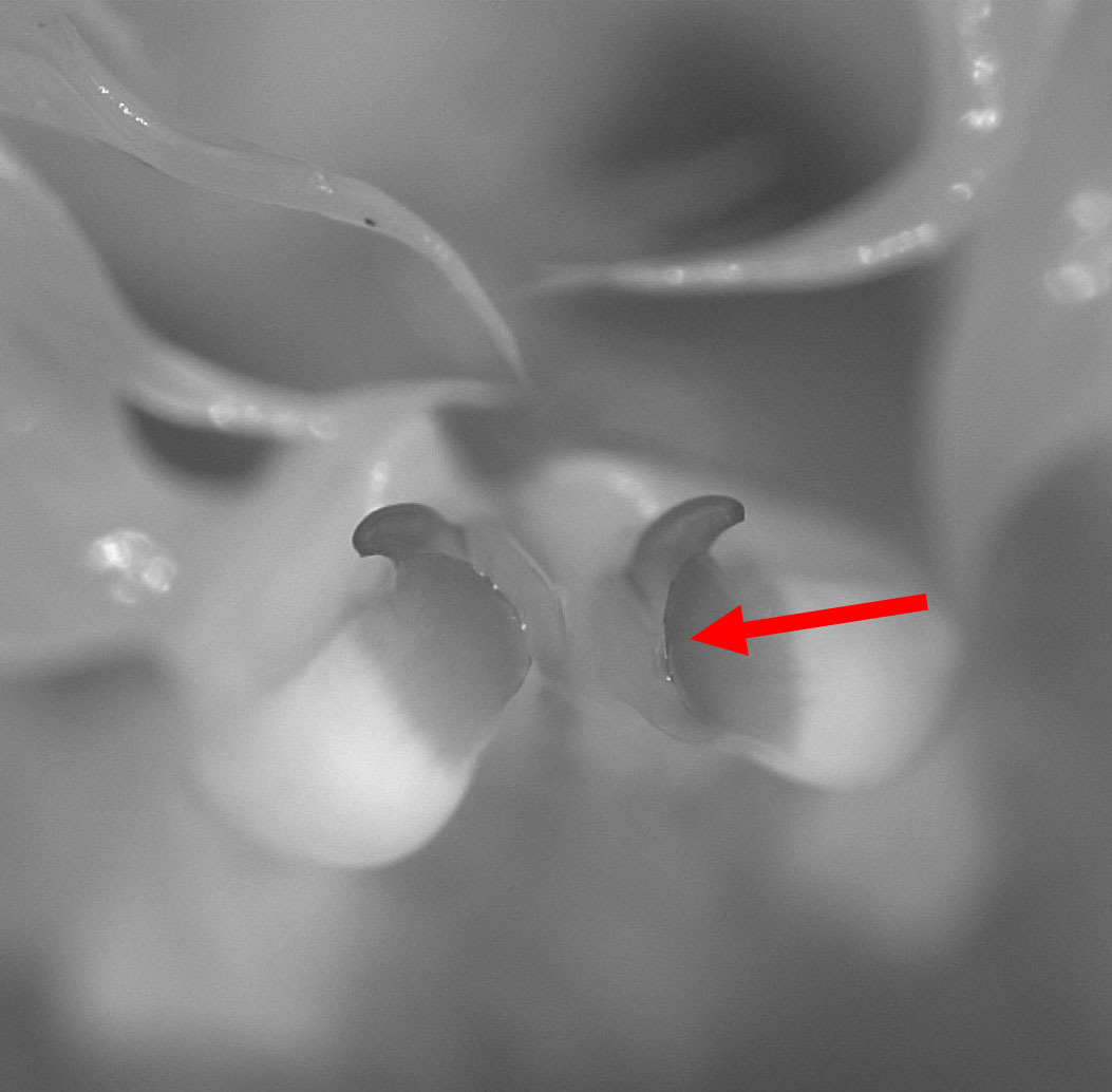
Below is a closeup of the apex of the gonopod of Procambarus simulans.
What is pointed to is the caudal process; in P. simulans it is flattened so
it's widest in this view:
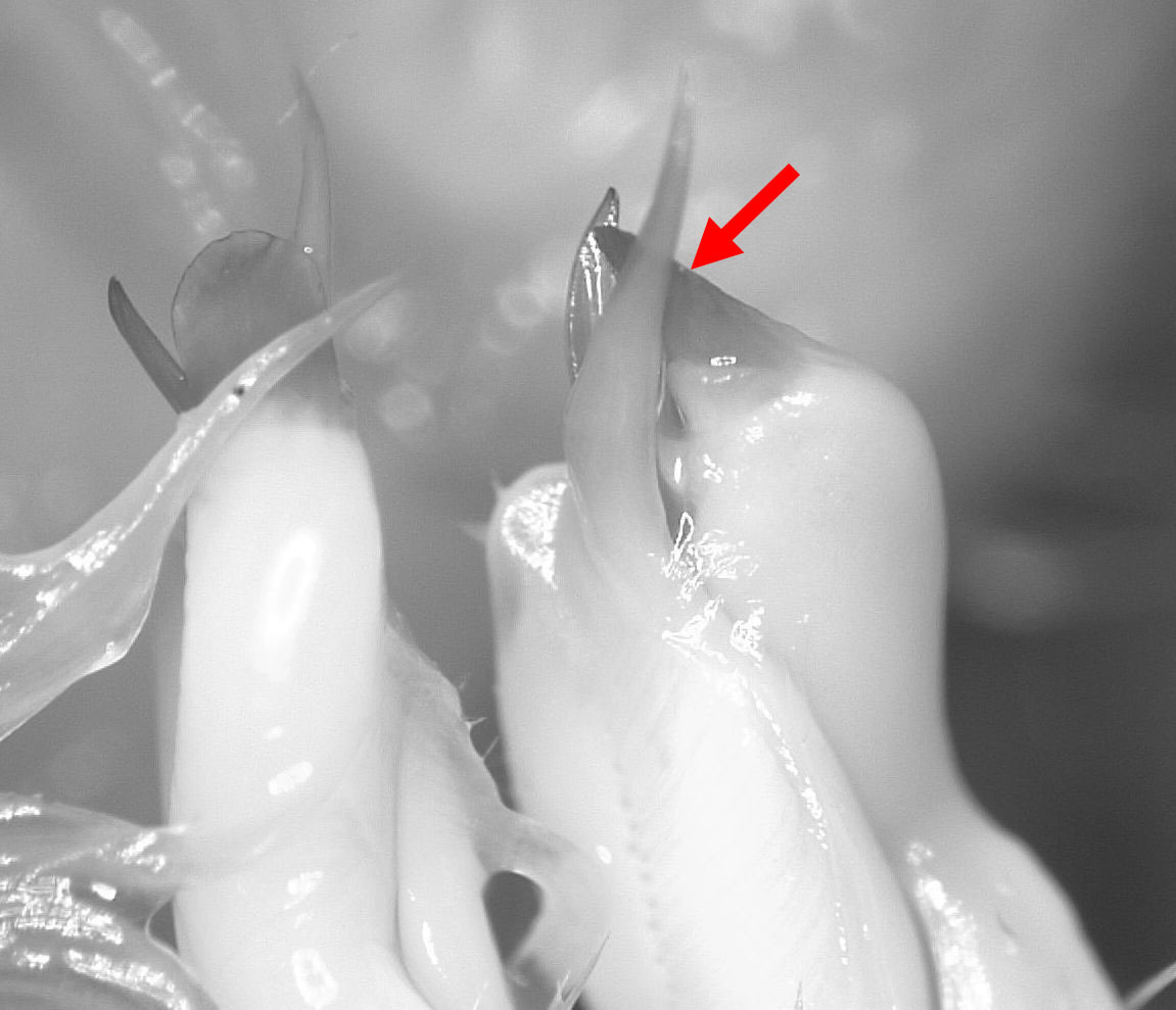
In contrast, the image below is from the same angle for Procambarus curdi. Here
the caudal process is flattened in a nearly perpendicular direction, so it appears narrow
in this view.
In the two images above, notice the right gonopod (one on left in photos). This same
process is in the perpendicular direction, so appears very narrow for P. simulans and very wide for P. curdi.
The above views should be sufficient. Another gonopod view that is alternatively be
useful is the "end on" view. Below is that view for P. simulans.
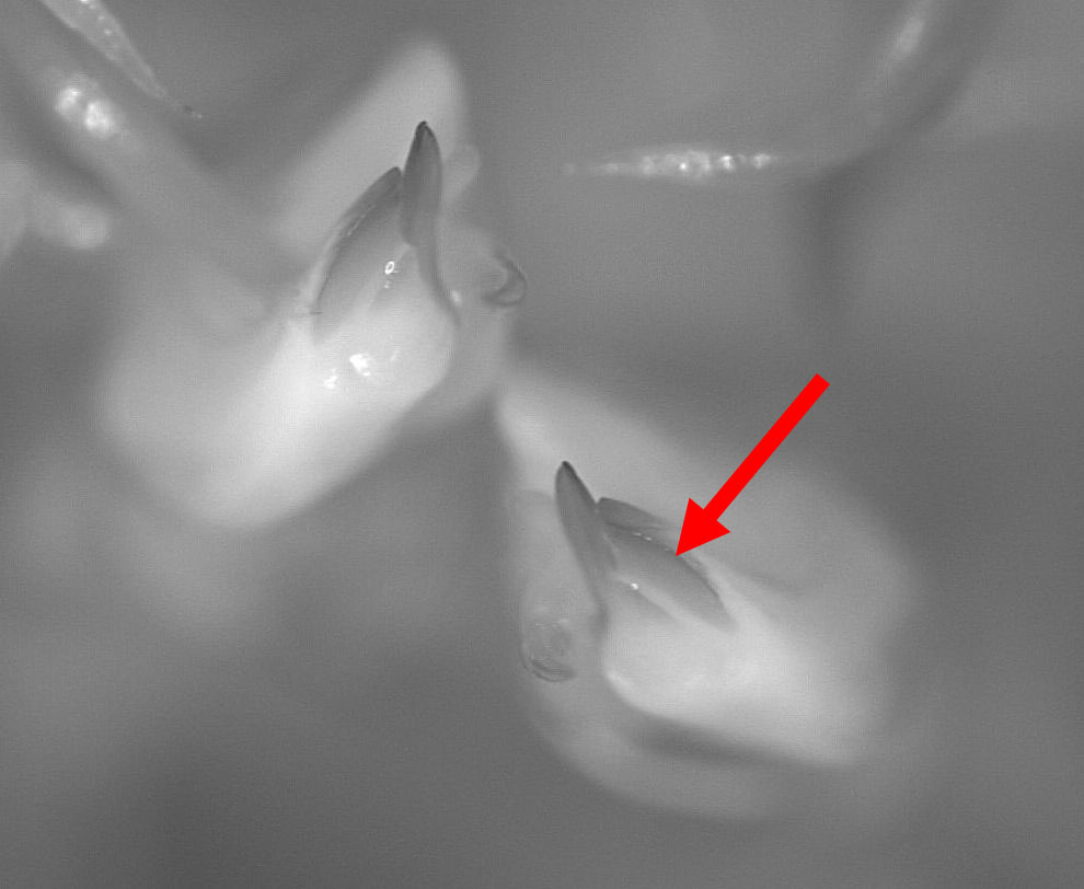
In constrast, the next image is for P. curdi. Notice the strikingly different
orientation of the caudal processes for the two species.
Males of these two species can be distinguished with such photos most easily
in adults, but also to a lesser degree in subadults.
Warning!!! Simply taking a photo of the region of the gonopods is almost never adequate. The
apices must be clearly visible. In many, many existing observations, the gonopods are
covered with hair, various debris, and often obscured by the 2nd pair of swimmerets
(gonopods are the first pair).
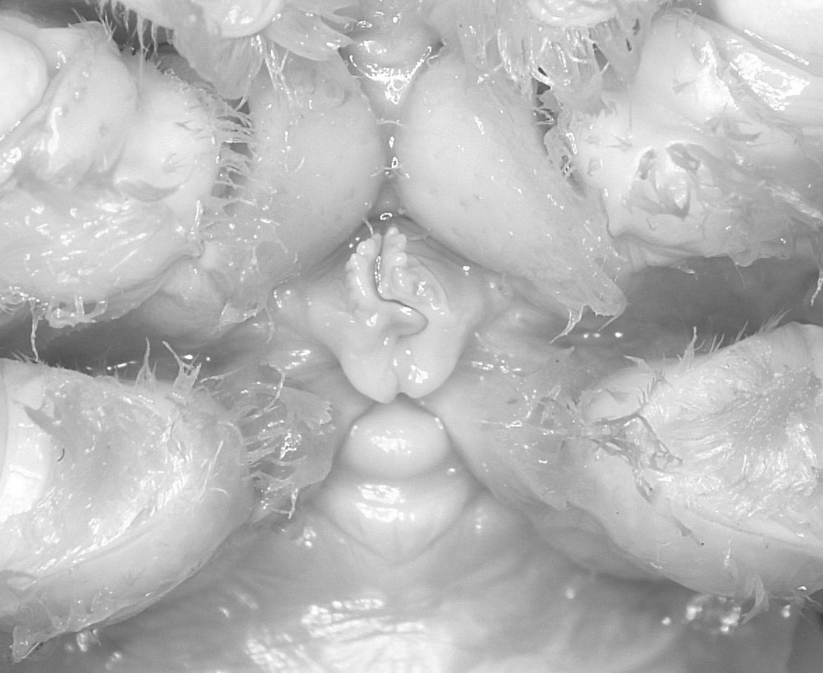
Females can be distinguished by different sinus shapes of the annulus ventralis
(sperm receptacle). Below
is a female P. curdi indicating the annulus ventralis.
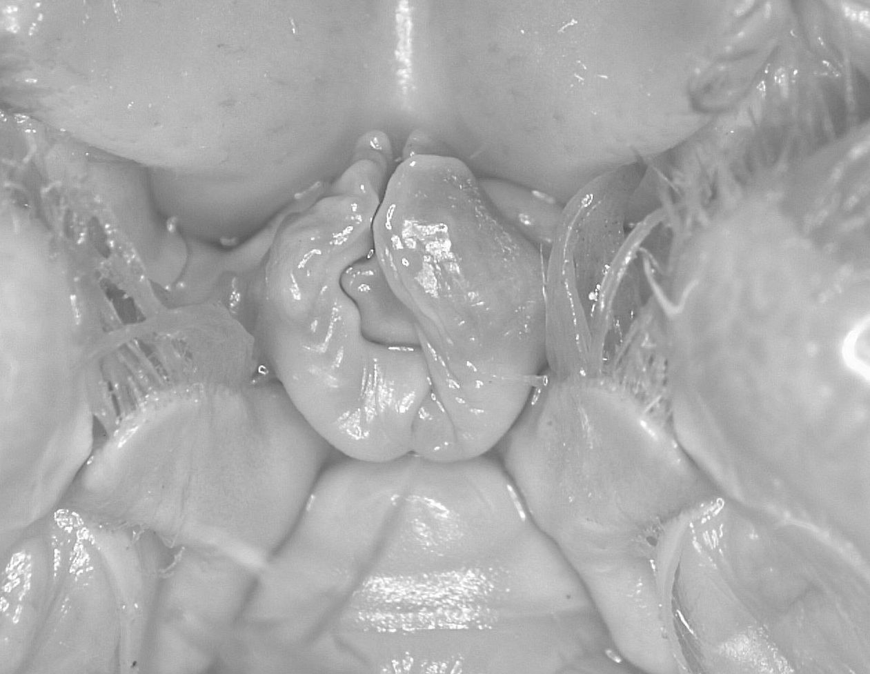
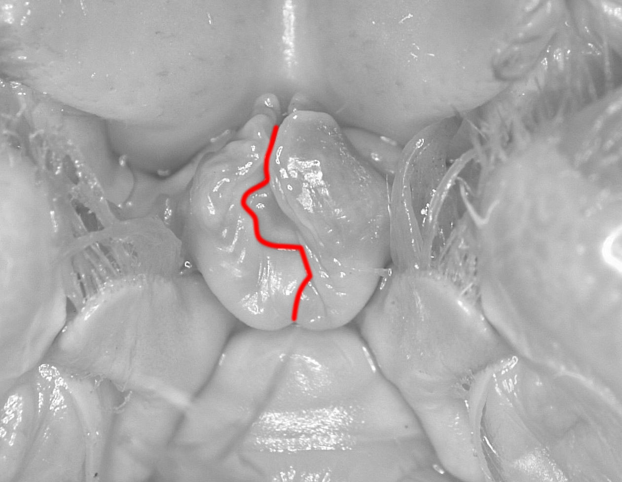
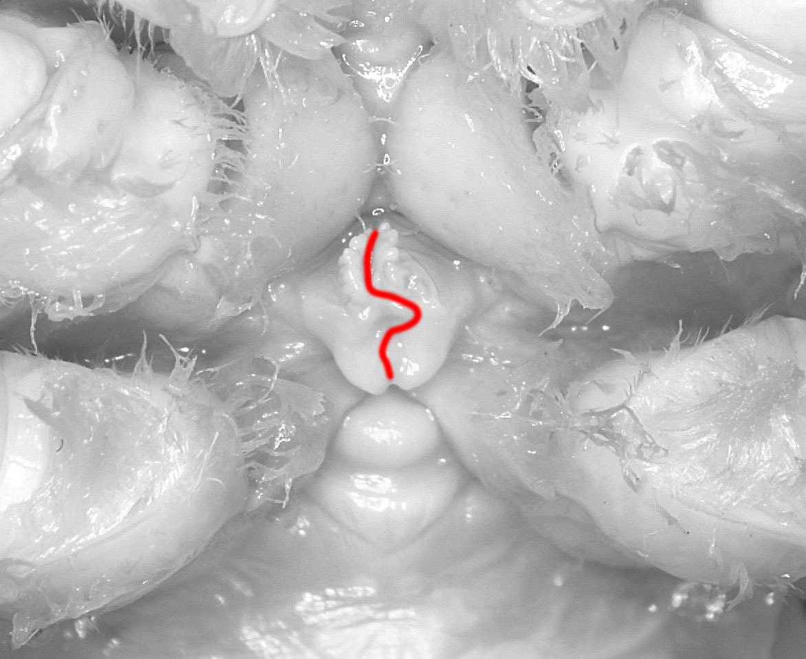
The two images below show a P. simulans annulus ventral without and with the sinus
highlighted:
In contrast, the pair of images below illustrate the sinus of P. curdi
Females of these two species can usually be distinguished by such photos for adult to
fairly small juvenile sizes.
Warning!!! Simply taking a photo of the ventral surface of the female, as is often done,
is rarely adequate for identification. The sinus of the annulus ventralis needs to
be clearly visible. In many existing photos it is obscured by hair and debris.


Comentarios
Añade un comentario