disc lichen
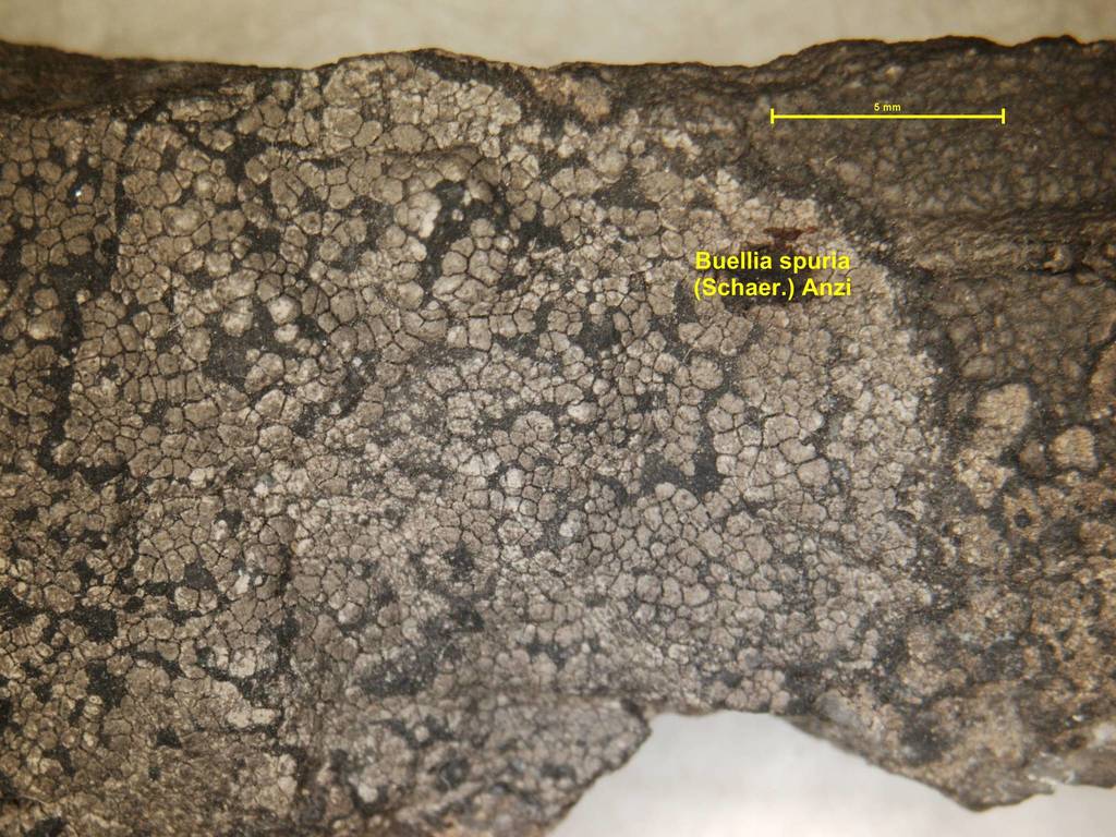
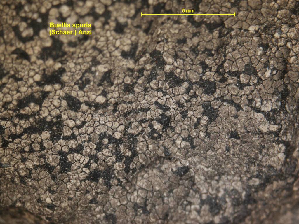
Buellia spuria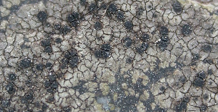

Description 6
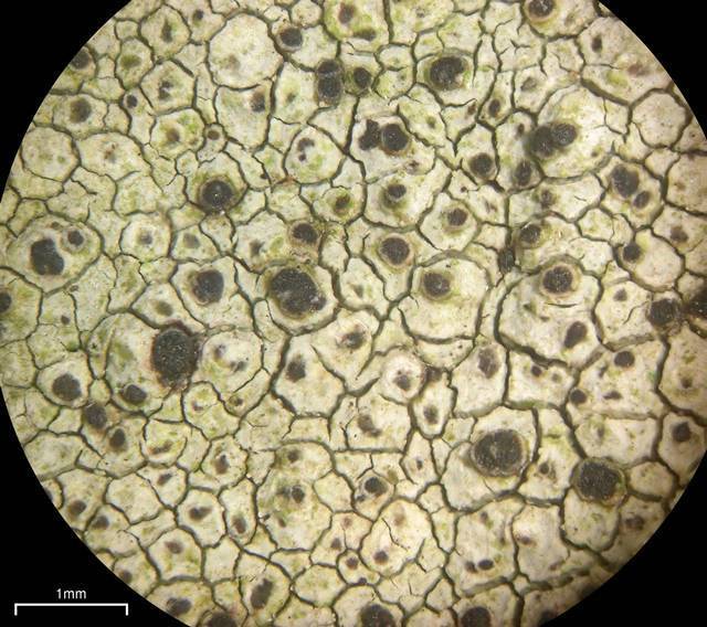
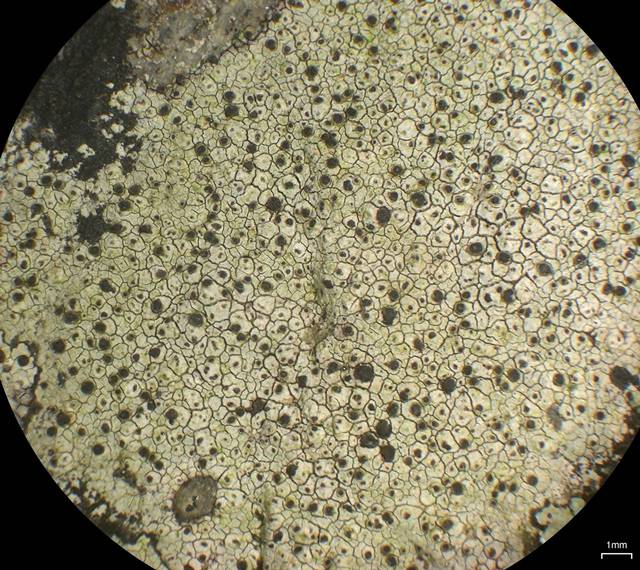
Thallus: crustose, areolate, thin to moderately thickened, ±continuous; prothallus: conspicuous, black, sometimes strongly developed and growing between the areoles (forming a hypothallus), rarely only surrounding the thallus outline; surface: usually white to whitish gray, rarely dark gray, dull or ±shiny, epruinose, phenocorticate, esorediate; medulla: white, lacking calcium oxalate (H2SO4-); Apothecia: lecideine; (0.2-)0.3-0.4(-0.5) mm in diam., immersed to adnate, rarely sessile; margin: black or color masked by grayish remains of necrotic thalline material (thalline veil), thin, ±persistent, rarely excluded with age; disc: black, epruinose, plane, rarely becoming slightly convex with age; proper exciple: narrow, poorly differentiated, aethalea-type, inner excipular hyphae narrow, hyaline, prosoplectenchymatous (textura oblita), often reduced, similar in structure and orientation to the paraphyses, transient with the deep reddish brown hypothecium (leptoclinoides-brown, textura intricata), outer excipular hyphae parallel, moderately swollen (textura oblita) and strongly carbonized with various amounts of brown and aeruginose pigments (cf. elachista-brown and cinereorufagreen, HNO3+ violet); epihymenium: brown, pigmentation continuous with the outer exciple (HNO3+ violet); hymenium: hyaline, not inspersed with oil droplets; paraphyses: simple to moderately branched, apically swollen, with a brown pigment cap (cf. elachista-brown); asci: clavate, Bacidia-type, 8-spored; ascospores: soon brown, 1-septate, oblong to ellipsoid, usually not constricted, with obtuse ends, not curved, (11-)11.9-[13.3]-14.8(-18) x (5-)5.9-[6.7]-7.4(-8) µm (n=60); proper septum: narrow, not thickening during spore ontogeny (Buellia-type); microrugulate or not visible in DIC; Pycnidia: rare, urceolate to globose, unilocular; ontogeny similar to the Umbilicaria-type; conidiogenous cells: mostly terminal, rarely also intercalary (cf. conidiophore-type V); conidia: bacilliform, 4.5-6 x 0.5-1 µm (n=20); Spot tests: usually K+ yellow to red (crystals), P- or + yellow, C-, KC-, CK-; fluorescence: UV- (dark); iodine reaction: thallus amyloid (only the medulla, not the cortex); Secondary metabolites: atranorin and chloroatranorin, norstictic, connorstictic, stictic with constictic, cryptostictic, hypostictic and methylstictic acids (J. A. Elix, HPLC).; Substrate and ecology: epilithic, on a variety of siliceous (HCl-) rock substrates, typically in montane habitats; World distribution: common and widely distributed throughout the Northern Hemisphere; Sonoran distribution: very common in Arizona, southern California, Baja California, Baja California Sur, Sonora, Chihuahua, and Sinaloa.; Notes: Buellia spuria is superficially most similar to B. stellulata, which has a different secondary chemistry and a thallus medulla that does react with iodine. Buellia spuria is widely distributed throughout the Sonoran Region, and, unlike B. stellulata, not restricted to oceanic regions. Some forms have mostly immersed apothecia, as does B. aethalea. Norstictic acid, a characteristic secondary metabolite of B. aethalea, is frequently also present in specimens of B. spuria, especially in material examined from the Sonoran Region. The K+ yellow to orange red spot test reaction can therefore not be used to distinguish the two species. However, TLC can easily distinguish all specimens because specimens of B. spuria contain atranorin and frequently stictic acid, neither found in B. aethalea. Apothecia of B. spuria are usually orbicular and not as strongly deformed as in B. aethalea. In most specimens of B. spuria, the apothecia eventually become sessile as opposed to remaining immersed as in B. aethalea.
Fuentes y créditos
- (c) André Aptroot, algunos derechos reservados (CC BY-NC-SA), http://www.tropicallichens.net/photopath/buellia-spuria-hongkong.jpg
- (c) Jason Hollinger, algunos derechos reservados (CC BY-SA), https://images.mushroomobserver.org/640/220517.jpg
- (c) Jason Hollinger, algunos derechos reservados (CC BY-SA), https://images.mushroomobserver.org/640/220518.jpg
- (c) National Museum of Natural History Collections, algunos derechos reservados (CC BY-NC-SA), https://collections.nmnh.si.edu/services/media.php?env=botany&irn=10522396
- (c) National Museum of Natural History Collections, algunos derechos reservados (CC BY-NC-SA), https://collections.nmnh.si.edu/services/media.php?env=botany&irn=10522445
- (c) Lichen Unlimited: Arizona State University, Tempe., algunos derechos reservados (CC BY-NC-SA), http://eol.org/data_objects/10549222


